Beranda
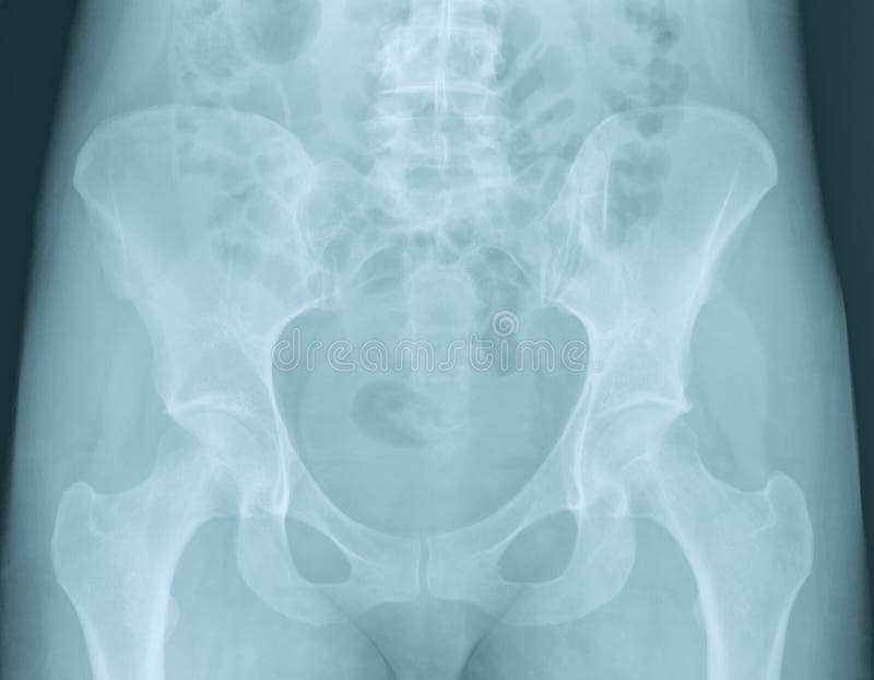
/ Pelvic Anatomy Xray : Pelvis Radiographic Anatomy - wikiRadiography / The bony pelvic girdle consists of the innominate bones bilaterally, and the sacrum and coccyx posteriorly.
Pelvic Anatomy Xray : Pelvis Radiographic Anatomy - wikiRadiography / The bony pelvic girdle consists of the innominate bones bilaterally, and the sacrum and coccyx posteriorly.
Insurance Gas/Electricity Loans Mortgage Attorney Lawyer Donate Conference Call Degree Credit Treatment Software Classes Recovery Trading Rehab Hosting Transfer Cord Blood Claim compensation mesothelioma mesothelioma attorney Houston car accident lawyer moreno valley can you sue a doctor for wrong diagnosis doctorate in security top online doctoral programs in business educational leadership doctoral programs online car accident doctor atlanta car accident doctor atlanta accident attorney rancho Cucamonga truck accident attorney san Antonio ONLINE BUSINESS DEGREE PROGRAMS ACCREDITED online accredited psychology degree masters degree in human resources online public administration masters degree online bitcoin merchant account bitcoin merchant services compare car insurance auto insurance troy mi seo explanation digital marketing degree floridaseo company fitness showrooms stamfordct how to work more efficiently seowordpress tips meaning of seo what is an seo what does an seo do what seo stands for best seotips google seo advice seo steps, The secure cloud-based platform for smart service delivery. Safelink is used by legal, professional and financial services to protect sensitive information, accelerate business processes and increase productivity. Use Safelink to collaborate securely with clients, colleagues and external parties. Safelink has a menu of workspace types with advanced features for dispute resolution, running deals and customised client portal creation. All data is encrypted (at rest and in transit and you retain your own encryption keys. Our titan security framework ensures your data is secure and you even have the option to choose your own data location from Channel Islands, London (UK), Dublin (EU), Australia.
Pelvic Anatomy Xray : Pelvis Radiographic Anatomy - wikiRadiography / The bony pelvic girdle consists of the innominate bones bilaterally, and the sacrum and coccyx posteriorly.. For more anatomy content please follow us and visit our website: Mri is a valuable technique in diagnosing or staging anomalies or conditions in the female pelvic region. (1) the iliopectineal line (2) the ilioishchial line (3) the dome of the acetabulum (4) the tear drop (5) the anterior rim of the acetabulum (6) the posterior rim of the acetabulum. It is of considerable importance in the management of severely injured patients presenting to emergency departments 1. Explore over 6700 anatomic structures and more than 670 000 translated medical labels.
Click here to load quiz. It is the most complete reference of human anatomy available on web, ipad, iphone and android devices. Mri anatomy images of the abdomen. Right pelvic renal transplant as seen on mra. This webpage presents the anatomical structures found on female pelvis mri.
Infographic Diagram Of Human Hip Bone Or Pelvic Girdle ... from media.istockphoto.com The pelvic spine consists of the sacrum and coccyx. It is the most complete reference of human anatomy available on web, ipad, iphone and android devices. Angiography invasive angiography is the gold standard modality for assessing pelvic vasculature 3. 1.5 retropubic anatomy showing points of attachments of the atla and the atfp. The spread of pelvic tumors to lymph nodes is an important means of tumor dissemination and substantially affects prognosis and management. Mri is a valuable technique in diagnosing or staging anomalies or conditions in the female pelvic region. Designed to help you quickly learn or review normal anatomy and confirm variants, imaging anatomy: Documents similar to systematic review of pelvical xray.
Axial, coronal, sagittal, and 3d reconstructions accompany highly accurate and detailed medical drawings, assisting you in making an accurate diagnosis.
Annotated case courtesy of dr phillip marsh, radiopaedia.org, rid: Mands thorough break down of this commonly used ed diagnostic the pelvic xr. It is the most complete reference of human anatomy available on web, ipad, iphone and android devices. Quizzes about radiology anatomy quiz. This webpage presents the anatomical structures found on female pelvis mri. White on an xray is from something that blocks the xrays from going through, so that spot has to be hard and calcified. The pelvic spine consists of the sacrum and coccyx. Figure 3a schematics show the anatomy of the female pelvic floor at the level of the pelvic diaphragm (a) and the urogenital diaphragm (b). For more anatomy content please follow us and visit our website: The main purposes of the pelvic girdle are to support and protect the abdominal and pelvic organs, and to connect the trunk and lower limbs. Documents similar to systematic review of pelvical xray. Click on the figure to see a description of each section. Click here to load quiz.
The pelvic diaphragm is composed of the ischiococcygeus muscle and levator ani muscle, the latter of which consists of the iliococcygeus, puborectalis, and pubococcygeus muscles. It is of considerable importance in the management of severely injured patients presenting to emergency departments 1. The pelvic spine consists of the sacrum and coccyx. Pelvis anatomy the pelvis is either the lower part of the trunk of the human body between the abdomen and the thighs. Figure 3a schematics show the anatomy of the female pelvic floor at the level of the pelvic diaphragm (a) and the urogenital diaphragm (b).
Pelvis Radiographic Anatomy - wikiRadiography from image.wikifoundry.com Biliary tract scan hida scan. Chest, abdomen, pelvis provides detailed views of anatomic structures in successive imaging slices in each standard plane of imaging. The pelvic anatomy tutorial was separated into four core sections: The spread of pelvic tumors to lymph nodes is an important means of tumor dissemination and substantially affects prognosis and management. The pelvic spine consists of the sacrum and coccyx. (1)the iliopectineal line is the radiographic landmark for the anterior column. Angiography invasive angiography is the gold standard modality for assessing pelvic vasculature 3. We think this is the most useful anatomy picture that you need.
Pelvis anatomy the pelvis is either the lower part of the trunk of the human body between the abdomen and the thighs.
The main purposes of the pelvic girdle are to support and protect the abdominal and pelvic organs, and to connect the trunk and lower limbs. Mri is a valuable technique in diagnosing or staging anomalies or conditions in the female pelvic region. Axial, coronal, sagittal, and 3d reconstructions accompany highly accurate and detailed medical drawings, assisting you in making an accurate diagnosis. Designed to help you quickly learn or review normal anatomy and confirm variants, imaging anatomy: At birth, each pelvic half consists of 3 separate primary bones: Use the mouse scroll wheel to move the images up and down alternatively use the tiny arrows (>>) on both side of the image to move the images.>>) on both side of the image to move the images. For more anatomy content please follow us and visit our website: White on an xray is from something that blocks the xrays from going through, so that spot has to be hard and calcified. Annotated case courtesy of dr phillip marsh, radiopaedia.org, rid: Annotated case courtesy of dr matthew lukies, radiopaedia.org, 51247. Angiography invasive angiography is the gold standard modality for assessing pelvic vasculature 3. The bony pelvic girdle consists of the innominate bones bilaterally, and the sacrum and coccyx posteriorly. Click here to load quiz.
1.5 retropubic anatomy showing points of attachments of the atla and the atfp. (1)the iliopectineal line is the radiographic landmark for the anterior column. Angiography invasive angiography is the gold standard modality for assessing pelvic vasculature 3. It includes several structures : Ct, mri, radiographs, anatomic diagrams and nuclear images.
Female Pelvis X-Ray Royalty Free Stock Photos - Image: 7064298 from thumbs.dreamstime.com Mands thorough break down of this commonly used ed diagnostic the pelvic xr. We think this is the most useful anatomy picture that you need. Right pelvic renal transplant as seen on mra. Angiography invasive angiography is the gold standard modality for assessing pelvic vasculature 3. Magnetic resonance imaging or mri of the female pelvis offers a unique display of the pelvic anatomy, including a woman's ovaries, uterus, and fallopian tubes. Conclusions • the primary imaging modalities for the abdomen and pelvis are plain film, ultrasound, and ct. (1)the iliopectineal line is the radiographic landmark for the anterior column. Chest, abdomen, pelvis provides detailed views of anatomic structures in successive imaging slices in each standard plane of imaging.
Right pelvic renal transplant as seen on mra.
The pelvic diaphragm is composed of the ischiococcygeus muscle and levator ani muscle, the latter of which consists of the iliococcygeus, puborectalis, and pubococcygeus muscles. Mri anatomy images of the abdomen. Quizzes about radiology anatomy quiz. It is of considerable importance in the management of severely injured patients presenting to emergency departments 1. This webpage presents the anatomical structures found on female pelvis mri. At birth, each pelvic half consists of 3 separate primary bones: During the secondary survey, pelvic bones are not stable, and there is a pain on palpation. The spread of pelvic tumors to lymph nodes is an important means of tumor dissemination and substantially affects prognosis and management. Figure 3a schematics show the anatomy of the female pelvic floor at the level of the pelvic diaphragm (a) and the urogenital diaphragm (b). Mri is a valuable technique in diagnosing or staging anomalies or conditions in the female pelvic region. Mands thorough break down of this commonly used ed diagnostic the pelvic xr. Documents similar to systematic review of pelvical xray. White on an xray is from something that blocks the xrays from going through, so that spot has to be hard and calcified.
The main purposes of the pelvic girdle are to support and protect the abdominal and pelvic organs, and to connect the trunk and lower limbs pelvic anatomy. The main purposes of the pelvic girdle are to support and protect the abdominal and pelvic organs, and to connect the trunk and lower limbs.